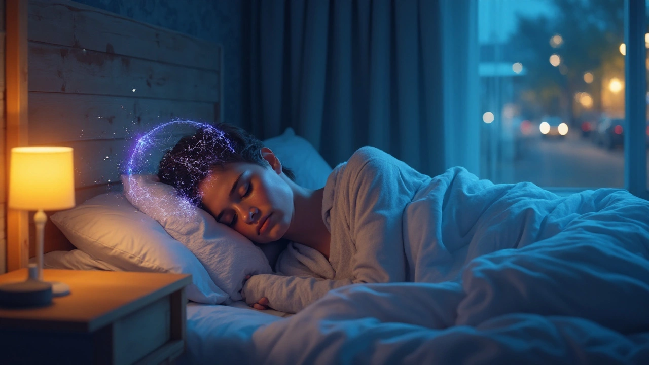
Sleep‑related partial onset seizures are episodes that begin in a specific brain region and are triggered or worsened by the sleep‑wake cycle. They represent a key intersection of sleep physiology and epilepsy dynamics, affecting millions worldwide.
What Is a Partial Onset Seizure?
Partial onset seizure (also called focal seizure) starts in a localized cortical area and may remain confined or spread to involve the whole brain. Typical features include sensory changes, motor jerks, or altered awareness, depending on the region involved. In clinical practice, these seizures account for roughly 60% of all epileptic events, according to the International League Against Epilepsy.
Why Sleep Matters for Epilepsy
Sleep is a reversible physiological state characterized by reduced consciousness, altered brainwave activity, and distinct stages (REM and NREM). During sleep, neuronal excitability shifts, making the brain either more or less prone to seizures. A 2023 longitudinal study of 1,200 epilepsy patients showed a 30% increase in seizure frequency during nights with fragmented NREM sleep.
Sleep Stages and Their Influence on Seizure Threshold
Two major sleep phases play opposite roles:
- REM sleep exhibits high acetylcholine levels and a desynchronized EEG pattern, which can lower the seizure threshold for some focal epilepsies.
- NREM sleep (especially stage N3, deep slow‑wave sleep) promotes neuronal inhibition, often protecting against seizures, though in certain focal syndromes it may paradoxically trigger events.
Understanding which stage drives a particular patient’s seizures guides both medication timing and sleep‑hygiene counseling.
Impact of Sleep Deprivation and Circadian Rhythm
Sleep deprivation is a well‑known seizure precipitant. A meta‑analysis (2022) of 15 trials found a 45% rise in seizure occurrence after 24‑hour sleep loss. The underlying mechanism involves up‑regulation of excitatory glutamate receptors and reduced GABAergic inhibition.
The circadian rhythm orchestrates hormone release (cortisol, melatonin) and ion channel expression. Disruptions-common in shift workers-can shift the peak seizure window from early morning to late night, complicating dose scheduling of antiepileptic drugs (AEDs).
Clinical Monitoring: EEG and Sleep Architecture
The gold standard for linking sleep to seizure activity is overnight electroencephalography (EEG). Video‑EEG captures both brainwave patterns and behavioral manifestations, revealing whether seizures cluster in REM, NREM, or occur independently of sleep.
Key EEG findings:
- Spike‑and‑wave discharges that intensify during NREM stage 2.
- Focal slowing that surges during REM, indicating heightened cortical excitability.
These patterns help neurologists adjust AED dosing (e.g., higher nighttime trough levels) or recommend targeted sleep‑intervention strategies.

Comparison of How Sleep Affects Seizure Types
| Attribute | Partial Onset (Focal) | Generalized |
|---|---|---|
| Typical Sleep Stage Trigger | Often NREM stage 2 (spike‑and‑wave bursts) | Predominantly REM for absence seizures |
| Response to Sleep Deprivation | Marked increase (≈40‑50%) | Variable; some tonic‑clonic seizures unaffected |
| EEG Signature During Sleep | Focal slowing, interictal spikes in NREM | Generalized 3‑Hz spike‑and‑wave in REM |
| Optimal AED Timing | Higher night‑time levels (e.g., extended‑release carbamazepine) | Evenly spaced dosing (e.g., valproate) |
Practical Strategies to Reduce Night‑Time Focal Seizures
- Maintain a consistent bedtime - aim for 7‑9hours of uninterrupted sleep.
- Limit caffeine and electronic screens after 6pm; blue‑light suppresses melatonin, which can destabilize neuronal firing.
- Use a sleep‑tracker or actigraphy to identify which sleep stage coincides with seizure spikes.
- Discuss with your neurologist the possibility of a once‑daily extended‑release AED to smooth nighttime drug levels.
- If nocturnal seizures persist, consider a bedside pulse‑oximeter or a seizure‑alert pillow to prompt early intervention.
These steps address both the physiological and behavioral contributors identified earlier.
Related Concepts and Next Steps
Understanding the link between sleep and focal seizures opens doors to other topics:
- Sleep‑related epileptiform discharges - how interictal spikes appear only during certain stages.
- Nocturnal epilepsy syndromes - rare conditions where seizures only occur at night.
- Chronotherapy - timing medication to the body’s circadian rhythm.
- Behavioral sleep medicine - CBT‑I for patients with epilepsy‑related insomnia.
Readers interested in deeper dives should explore these adjacent areas, which together form a comprehensive knowledge cluster around epilepsy and sleep health.
Key Takeaway
The interplay between sleep and partial onset seizures is intricate but manageable. By recognizing which sleep stages exacerbate seizures, monitoring with overnight EEG, and applying disciplined sleep‑hygiene, patients can often lower seizure frequency and improve overall quality of life.
Frequently Asked Questions
Can I have a seizure while dreaming?
Yes. Seizures that start in REM sleep often occur when the brain is active in a dreaming state. They may manifest as motor jerks or visual hallucinations that the sleeper perceives as part of the dream.
Is it safe to skip a night’s sleep if I’m on medication?
Skipping sleep dramatically raises seizure risk, even when AED levels are therapeutic. A single night of total sleep deprivation can increase focal seizure frequency by up to 45%.
Do all AEDs work better at night?
Not all. Drugs with long half‑lives (e.g., lamotrigine) maintain stable levels, while short‑acting agents (e.g., carbamazepine) may benefit from nighttime dosing to cover the sleep period when seizures peak.
How does a sleep‑tracker help me?
A reliable tracker can flag prolonged NREM periods or fragmented REM cycles. Correlating these logs with seizure diaries lets you see patterns and discuss them with your neurologist.
Are there surgical options for sleep‑triggered focal seizures?
When seizures are localized to a resectable region and remain uncontrolled despite optimal medication and sleep management, epilepsy surgery (e.g., focal cortical resection) may be considered. Pre‑surgical video‑EEG monitoring often includes sleep to map the exact trigger.


sleep deprived for 3 days straight last month tried to fix my circadian rhythm and ended up having 4 seizures in one night guess who's not sleeping anymore